News
Laryngeal paralysis
8th August 2022
Laryngeal paralysis is a well- documented cause of obstruction of the upper portion of the respiratory tract in horses, dogs, and humans but it is considered uncommon in cats.
Laryngeal paralysis can be unilateral or bilateral, congenital or acquired and it results from the failure in abduction of one or both arytenoid cartilages which leads to a narrowing of the rima glottis. In many patients the cause remains undetermined, and these cases were traditionally classified as idiopathic; it has recently been shown that many of these dogs with acquired laryngeal paralysis develop systemic neurologic signs within one year following diagnosis of laryngeal paralysis, which is consistent with a progressive generalized neuropathy.
Dogs typically present with noisy inspiratory respiration and exercise intolerance. Early clinical signs include voice change and mild coughing and gagging. Severe airway obstruction results in respiratory distress, cyanosis, and collapse. Dogs can also show dysphagia or develop rear limb weakness associated with peripheral neuropathy. The classic finding on physical examination is the presence of stridor over the upper airway, but this can be variable. The clinical signs are often life-threatening in cats and affect those patients suffering from both unilateral and bilateral laryngeal paralysis.
Routine diagnostic evaluation for patients thought to have laryngeal paralysis includes physical examination, orthopaedic and neurologic examination, complete blood count, biochemical profile, urinalysis, thyroid function screening, thoracic radiographs, and laryngeal examination. Thoracic radiographs are a necessary part of the diagnostic work-up in dogs suspected to have laryngeal dysfunction to rule out not only aspiration pneumonia but also overt megaesophagus, pulmonary oedema, and concurrent cardiac or lower airway abnormalities. Hypothyroidism occurs concurrently in approximately 30% of dogs with acquired laryngeal paralysis, although a direct causal link has not been established. Regardless, thyroid function screening is performed routinely in the work-up for laryngeal paralysis. Thyroid supplementation should be instituted if indicated, although this does not resolve clinical signs associated with laryngeal paralysis. Laryngeal examination under a light plane of anaesthesia is required to provide a definitive diagnosis of laryngeal paralysis and to rule out other laryngeal abnormalities. This examination can be accomplished by direct visualization of the larynx with a simple laryngoscope, oral video-endoscopic laryngoscopy, transnasal laryngoscopy (TNL), ultrasonography (echolaryngography), or computed tomography (CT). Laryngeal paralysis is diagnosed based on the lack of arytenoid abduction during inspiration. Inflammation and swelling of the laryngeal cartilages can also be apparent. Diagnosis can be confounded by paradoxic movement of the arytenoids, resulting in a false-negative result. In this scenario, the arytenoid cartilages move inward during inspiration because of negative intraglottic pressure created by increased respiratory effort against an obstruction. The cartilages then passively return to a normal position during expiration, which gives the impression of normal arytenoid movement. To avoid this situation, an assistant should state the phase of ventilation during laryngoscopy to distinguish normal from abnormal motion.
Laryngeal paralysis is a surgical condition for severely affected dogs (bilateral laryngeal paralysis) and for all the cats. The surgical treatments reported are various: arytenoid cartilage lateralization (unilateral or bilateral), partial laryngectomy, castellated laryngofissure, permanent tracheostomy, ventriculocordectomy. Unilateral arytenoid lateralization is co
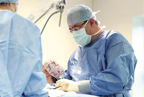
nsidered the procedure of choice in dogs with a low complication rate and good to excellent outcomes.
The aim of the procedure in dogs and cats is to increase the area of the rima glottis. Modifications of this surgical technique have been reported in the dog: cricoarytenoid lateralization (CAL), thyroarythenoid lateralization (TAL), with or without cricothyroid, cricoarytenoid and interarytenoid disarticulation. Numerous complications have been reported with these surgical techniques, including aspiration pneumonia, laryngeal webbing, surgical failure, seroma formation at the surgical site, airway reobstruction secondary to laryngeal edema, and transient Horner’s syndrome.
In the absence of surgical complications, unilateral arytenoid lateralization results in reduced respiratory distress and stridor and improved exercise tolerance. Owner satisfaction with this procedure is excellent, with most owners believing that the quality of the dog’s life was improved dramatically.
Back to news & events